Effect of Different Scan Range for CT Imaging on Stress Distribution in Implant and peripheral bone
Dental Implant And Stress Analysis
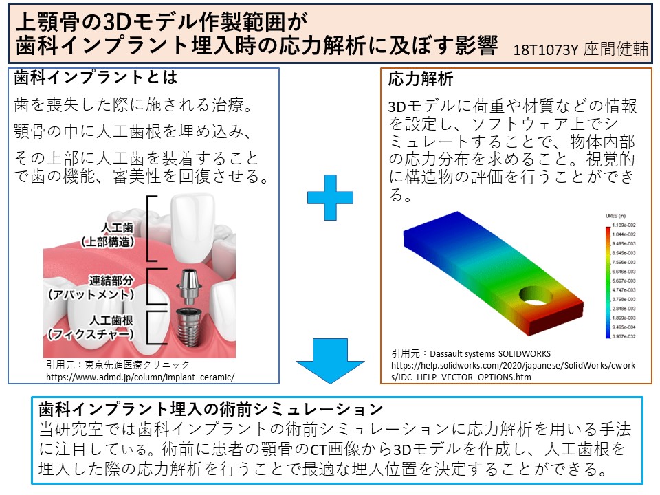
Dental implant treatment is one of the treatments for tooth loss.
An artificial tooth root is implanted in the jawbone and covered with an artificial tooth to restore dental function and esthetics.
Our laboratory is researching a method to create a 3D model of a patient's jawbone and analyze the stress during dental implant placement as a preoperative simulation of dental implant surgery. This method enables us to verify the load on the bone in advance and to determine the optimal placement position.
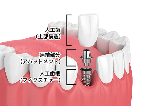
Schematic of Dental Implants[1]
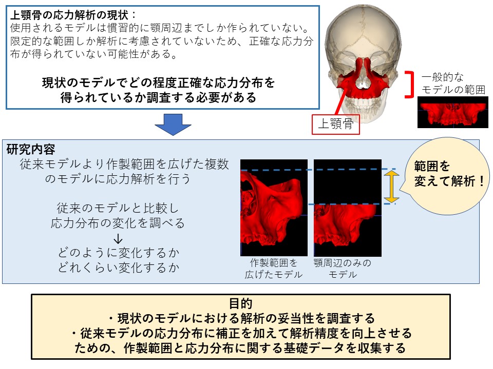
The 3D model used for stress analysis is created based on CT images taken for each patient. In the case of the maxilla, the model used in the analysis is conventionally created only up to the jaw area. However, since the maxilla is part of a larger bone, the skull, only a limited portion of the bone is considered in this model. In addition, there is a lack of data on the analysis of models with a wider range of fabrication, so it is not known how accurate the analysis is with the current model.
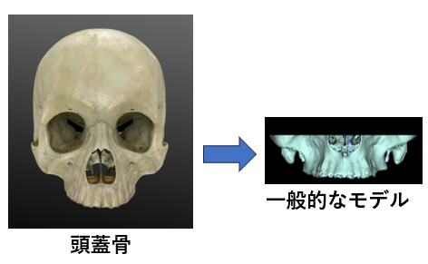
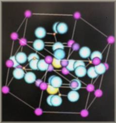
3D model of maxilla
Objective
To investigate how much the stress distribution in the maxilla is affected by the different ranges of the 3D model, and to verify the validity of the analysis on the current model.
Collect data showing the relationship between the fabrication area and stress distribution in order to improve the accuracy of analysis with conventional models.
Slides
[1]東京先進医療クリニック
https://www.admd.jp/column/implant_ceramic/